
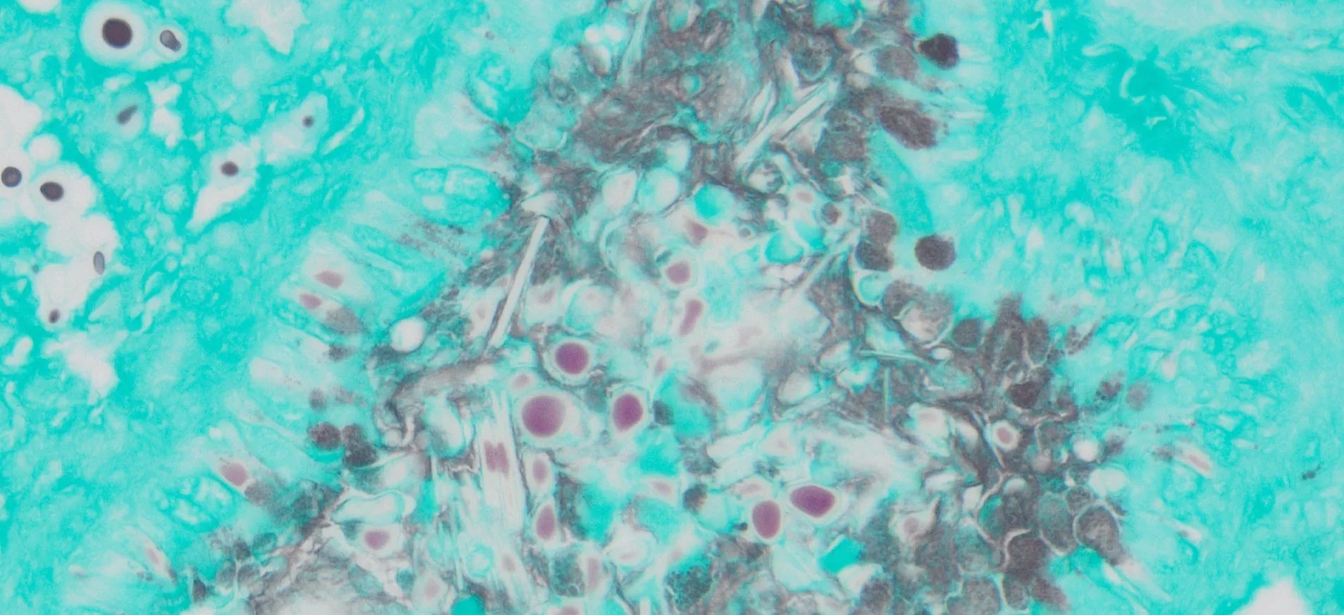
Tissue-based infection research becomes more informative when investigators can see both the organism and the surrounding cellular response. Methenamine silver staining is useful for highlighting fungal elements in FFPE or frozen tissue sections, allowing researchers to map where fungal structures are located within preserved morphology.
Localization alone does not explain how nearby cells are responding. Tissue injury may involve inflammation, structural disruption, or DNA fragmentation, so complementary methods can help researchers examine separate biological events without expecting one stain to answer every question.
Start With Spatial Evidence
A fungal stain provides an anatomical map of where organisms are present. Grocott-Gomori methenamine silver chemistry oxidizes mucopolysaccharides in fungal cell walls and produces a dark metallic-silver reaction, creating strong contrast against the surrounding tissue.
Researchers can then examine adjacent regions for evidence of cellular damage. A tunel assay kit detects DNA breaks associated with extensive DNA degradation during late-stage apoptosis, providing a separate readout that can be compared with fungal distribution rather than interpreted as direct proof of infection.
Keep the Two Endpoints Separate
Fungal visualization and TUNEL labeling measure different biological features. A black-stained fungal structure identifies the location of fungal material, while a TUNEL-positive nucleus indicates substantial DNA fragmentation in a cell.
The two signals should therefore be analyzed independently first. Researchers can then ask whether areas containing fungal elements also show increased DNA fragmentation, whether changes occur at a distance, or whether different tissue compartments respond differently.
Use Adjacent Sections Strategically
Serial sections can help connect these observations without forcing incompatible staining chemistries onto one slide. One section can be reserved for fungal identification, while a neighboring section is prepared for TUNEL labeling under conditions that preserve the relevant DNA ends.
Because consecutive sections are not identical, investigators should record cutting level and anatomical landmarks. Small lesions or focal fungal structures can shift between levels, so careful region matching is important when comparing patterns across slides.
Protect Sample Quality During Preparation
GMS staining can be performed on paraffin or frozen sections, but the workflow includes oxidation, silver impregnation, toning, counterstaining, dehydration, and clearing. Consistent timing and reagent preparation are important for maintaining readable contrast.
TUNEL preparation follows a different path. Paraffin sections require deparaffinization and controlled permeabilization, while frozen sections need suitable fixation and handling before labeling. Excessive pretreatment can damage morphology, whereas insufficient treatment may reduce access to fragmented DNA.
Build Controls Into Both Assays
Positive controls are essential for confirming that each method is functioning. A fungus-containing tissue section can verify the expected dark fungal signal in GMS staining, while a known TUNEL-positive control confirms that DNA-break labeling is working.
Negative controls are equally useful for the TUNEL workflow because they help identify background caused by nonspecific detection. Processing controls alongside experimental slides makes it easier to separate technical problems from genuine biological variation.
Standardize Imaging and Region Selection
Microscope settings should remain consistent when researchers compare staining across groups. Magnification, illumination, exposure, white balance, and digital processing can all change the apparent strength or visibility of a signal.
Regions of interest should also be defined before full analysis begins. Investigators may compare fungal burden, TUNEL-positive cell counts, distance from fungal structures, or region-specific patterns, but the same selection and scoring rules should be applied to every specimen.
Avoid Overinterpreting TUNEL Positivity
DNA fragmentation can be associated with apoptosis, but TUNEL signal should still be interpreted with morphology, controls, and experimental context. A positive label does not by itself identify the cause of cell damage or prove that fungal material triggered the event.
Combining TUNEL with routine histology or additional molecular markers can strengthen interpretation. The goal is to build several independent lines of evidence rather than assigning a biological mechanism from one staining result.
Plan the Study as One Workflow
The strongest design begins before sectioning. Researchers should decide which block levels will be used, how slides will be allocated, which controls are needed, and how image analysis will connect organism localization with cellular-response measurements.
This planning protects limited material and reduces repeat work. It also makes it easier to preserve comparable sections for future assays if the first results reveal a new question about inflammation, cell identity, or tissue remodeling.
Conclusion
Pathogen-focused histology becomes more informative when organism localization is studied alongside carefully selected indicators of tissue response. Fungal staining and DNA-fragmentation analysis provide different layers of information that can be compared within the same anatomical context.
By keeping the assays technically separate, using adjacent sections, standardizing imaging, and applying suitable controls, researchers can build a clearer picture of how fungal presence relates to cellular change while avoiding conclusions that extend beyond what each method actually measures.